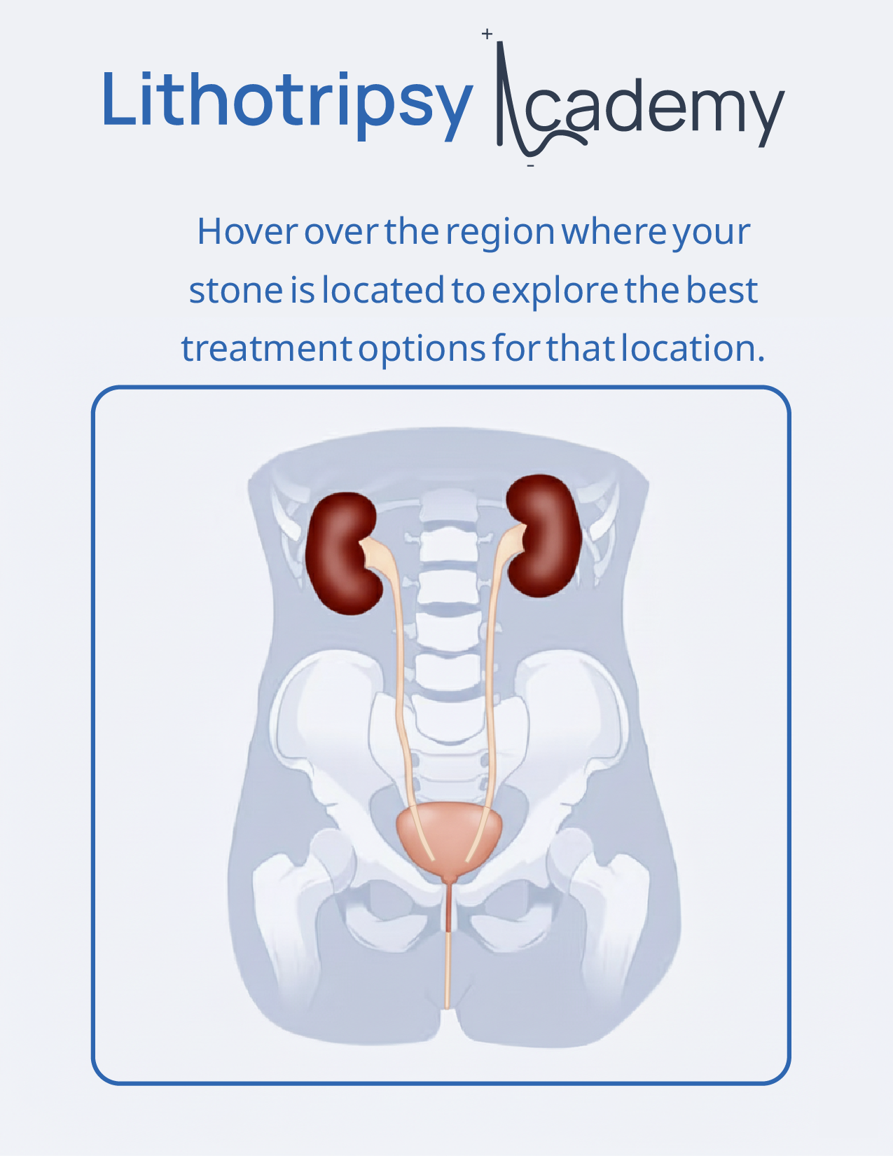
Understanding the basics of renal stones

Expert information, personalized support, and care you can trust — at every step.​

What are kidney stones?

Kidney stones, also known as renal stones or nephrolithiasis, are hard deposits made of minerals and salts that form inside your kidneys. These stones develop when your urine contains more crystal-forming substances—such as calcium, oxalate, and uric acid—than the fluid in your urine can dilute.

Urinary system anatomy
Common types of kidney stones:
  • Calcium Oxalate Stones: The most common type, formed when calcium combines with oxalate in the urine.
  • Uric Acid Stones: These form in people who lose too much fluid or eat a high-protein diet. They’re more common in individuals with gout.
  • Struvite Stones: Often develop after urinary tract infections and can grow quickly with few symptoms.
  • Cystine Stones: Rare and usually caused by a genetic disorder where the kidneys excrete too much of the amino acid cystine.

Symptoms to watch For:

  • Severe pain in the side and back, below the ribs
  • Pain that radiates to the lower abdomen and groin
  • Pain during urination
  • Pink, red, or brown urine
  • Nausea and vomiting
  • Urgent or frequent urination

Causes and Risk Factors:

Several factors can contribute to the formation of kidney stones, and the exact cause may vary based on the stone type. Some common causes and risk factors include:

Diet

Diets high in sodium, oxalate, and animal protein can increase the risk of stone formation.

Medication

Some medications, such as certain diuretics and antacids, can increase the likelihood of kidney stone development.

Family History

A family history of kidney stones may increase an individual's likelihood of developing stones.

Dehydration

Insufficient fluid intake can lead to concentrated urine, making it more likely for minerals and salts to crystallize and form stones.

Medical Conditions

Certain medical conditions, such as urinary tract infections, hyperparathyroidism, and gout, can increase the risk of stone formation.

Obesity

Being overweight or obese is associated with an increased risk of kidney stones.

Geographical Factors

Certain geographic regions with high temperatures and low humidity may contribute to dehydration and increase the risk of stone formation.

Previous History of Kidney Stones

People who have had kidney stones in the past are at a higher risk of experiencing them again.

Age and Gender

Men are more prone to kidney stones than women, and the risk generally increases with age.

Diagnosis of Urolithiasis

If you suspect you might have kidney stones, it’s important to see your doctor for a proper diagnosis. Kidney stones can cause significant pain, and identifying them is the first step towards getting relief and preventing future problems. Your doctor will likely use a combination of the following methods to determine if you have urolithiasis.

Medical History and Physical Examination:
Your doctor will start by asking you about your symptoms, including the location and severity of your pain, any nausea or vomiting, and your urine habits. They will also inquire about your past medical history, including any previous kidney stones, family history of kidney stones, dietary habits, and fluid intake. A physical examination may also be performed.

Urine Tests (Urinalysis):
Analyzing a sample of your urine can provide important clues. The presence of blood (hematuria), crystals, or signs of infection can suggest a kidney stone. Your doctor may also order a 24-hour urine collection to measure the levels of certain minerals and substances in your urine, which can help identify the underlying cause of stone formation and guide preventive measures.

Imaging Tests:
Imaging tests allow doctors to visualize your kidneys and urinary tract to confirm the presence of stones, determine their size and location, and identify any blockages. Common imaging techniques include:

  • Kidney, Ureter, and Bladder X-ray (KUB): This is a plain X-ray that can sometimes show larger, calcium-containing stones. However, smaller stones and certain types of stones may not be visible on a KUB.
  • Ultrasound: This non-invasive test uses sound waves to create images of your kidneys and bladder. It’s particularly useful for detecting stones and blockages, and it doesn’t involve radiation.
  • Computed Tomography (CT) Scan: A CT scan provides detailed cross-sectional images of your urinary tract. It is often the most effective way to identify even small stones and assess their exact location and size. A special type of CT scan called a “low-dose CT” can minimize radiation exposure. Intravenous Pyelogram (IVP): This involves injecting a contrast dye into a vein and then taking a series of X-rays. The dye highlights the kidneys and ureters, helping to identify blockages caused by stones. While effective, it’s used less frequently now due to the availability of CT scans.

Blood Tests:
Blood tests can help assess your kidney function and measure levels of certain minerals in your blood that may contribute to stone formation, such as calcium, phosphorus, and uric acid.
Based on the results of these tests, your doctor will be able to confirm a diagnosis of urolithiasis and determine the best course of treatment for you.

Treatment of Urolithiasis

The treatment for kidney stones depends on several factors, including the size, location, and type of stone, as well as the severity of your symptoms and overall health. Not all kidney stones require active treatment, as some small stones may pass on their own. However, if you are experiencing significant pain, have a blockage, or your stone is unlikely to pass naturally, medical intervention may be necessary.

Here are some common treatment options for urolithiasis:

Conservative Management (Watchful Waiting):
  • Pain Management: Using over-the-counter or prescription pain relievers to manage the discomfort.
  • Increased Fluid Intake: Drinking plenty of water (at least 2-3 liters per day) to help flush the stone out naturally.
  • Medications: In some cases, medications called alpha-blockers may be prescribed to relax the muscles in the ureter, making it easier for the stone to pass.
  • Strain Your Urine: Your doctor may advise you to strain your urine to collect any passed stones for analysis, which can help determine their composition and guide future prevention strategies.

ESWL is a non-invasive procedure that uses shock waves to break kidney stones into smaller pieces. These smaller fragments can then pass more easily through the urinary tract…read more  

If ESWL is not suitable or unsuccessful, other minimally invasive surgical options are available:

  • Ureteroscopy: This procedure involves passing a thin, flexible telescope with a camera (ureteroscope) through the urethra and bladder into the ureter. Once the stone is located, it can be retrieved with a small basket or forceps, or it can be broken into smaller pieces using a laser or other energy source (laser lithotripsy). This is often used for stones in the ureter or lower kidney.
  • Percutaneous Nephrolithotomy (PCNL): This procedure is typically used for larger kidney stones. A small incision is made in your back, and a hollow tube is inserted directly into the kidney. The stone is then located and removed or broken up using specialized instruments. PCNL usually requires a short hospital stay.

Treatment options

Hover over the region where your stone is located to explore the best treatment options for that location.

Treatment options
Renal Pelvis stone (right) Upper pole stone (right) Lower pole stone (right) Middle Pole Stone (right) Upper Ureter stone (right) Renal Pelvis stone (left) Upper pole stone (left) Middle Pole Stone (left) Lower pole stone (left) Upper Ureter stone (left) Lower Ureter stone (right) Lower Ureter stone (left)

Renal Pelvis stone (right)

If your stone is not larger than 15 mm and its density is below 1000 Hounsfield units, Shock Wave Lithotripsy (SWL) is often the best treatment option. It’s a non-invasive procedure, so you usually don’t need surgery.

Upper pole stone (right)

If your stone is smaller than 15 mm and has a density below 1000 Hounsfield units, Shock Wave Lithotripsy (SWL) is often a good treatment choice — as long as the anatomy is suitable. It’s a gentle, non-invasive procedure, so surgery is usually not necessary.

Lower pole stone (right)

If your stone is smaller than 15 mm and has a density below 1000 Hounsfield units, Shock Wave Lithotripsy (SWL) is often a good treatment choice — as long as the anatomy is suitable. It’s a gentle, non-invasive procedure, so surgery is usually not necessary.

Middle Pole Stone (right)

If your stone is smaller than 15 mm and has a density below 1000 Hounsfield units, Shock Wave Lithotripsy (SWL) is often a good treatment choice — as long as the anatomy is suitable. It’s a gentle, non-invasive procedure, so surgery is usually not necessary.

Upper Ureter stone (right)

If your stone is smaller than 15 mm and has a density below 1000 Hounsfield units, Shock Wave Lithotripsy (SWL) is often a good treatment choice — as long as the anatomy is suitable and the stone is not obstructing for long time. It’s a gentle, non-invasive procedure, so surgery is usually not necessary.

Renal Pelvis stone (left)

If your stone is not larger than 15 mm and its density is below 1000 Hounsfield units, Shock Wave Lithotripsy (SWL) is often the best treatment option. It’s a non-invasive procedure, so you usually don’t need surgery.

Upper pole stone (left)

If your stone is smaller than 15 mm and has a density below 1000 Hounsfield units, Shock Wave Lithotripsy (SWL) is often a good treatment choice — as long as the anatomy is suitable. It’s a gentle, non-invasive procedure, so surgery is usually not necessary.

Middle Pole Stone (left)

If your stone is smaller than 15 mm and has a density below 1000 Hounsfield units, Shock Wave Lithotripsy (SWL) is often a good treatment choice — as long as the anatomy is suitable. It’s a gentle, non-invasive procedure, so surgery is usually not necessary.

Lower pole stone (left)

If your stone is smaller than 15 mm and has a density below 1000 Hounsfield units, Shock Wave Lithotripsy (SWL) is often a good treatment choice — as long as the anatomy is suitable. It’s a gentle, non-invasive procedure, so surgery is usually not necessary.

Upper Ureter stone (left)

If your stone is smaller than 15 mm and has a density below 1000 Hounsfield units, Shock Wave Lithotripsy (SWL) is often a good treatment choice — as long as the anatomy is suitable and the stone is not obstructing for long time. It’s a gentle, non-invasive procedure, so surgery is usually not necessary.

Lower Ureter stone (right)

If your stone is smaller than 15 mm and has a density below 1000 Hounsfield units, Shock Wave Lithotripsy (SWL) is often a good treatment choice — as long as the anatomy is suitable and the stone is not obstructing for long time. It’s a gentle, non-invasive procedure, so surgery is usually not necessary.

Lower Ureter stone (left)

If your stone is smaller than 15 mm and has a density below 1000 Hounsfield units, Shock Wave Lithotripsy (SWL) is often a good treatment choice — as long as the anatomy is suitable and the stone is not obstructing for long time. It’s a gentle, non-invasive procedure, so surgery is usually not necessary.